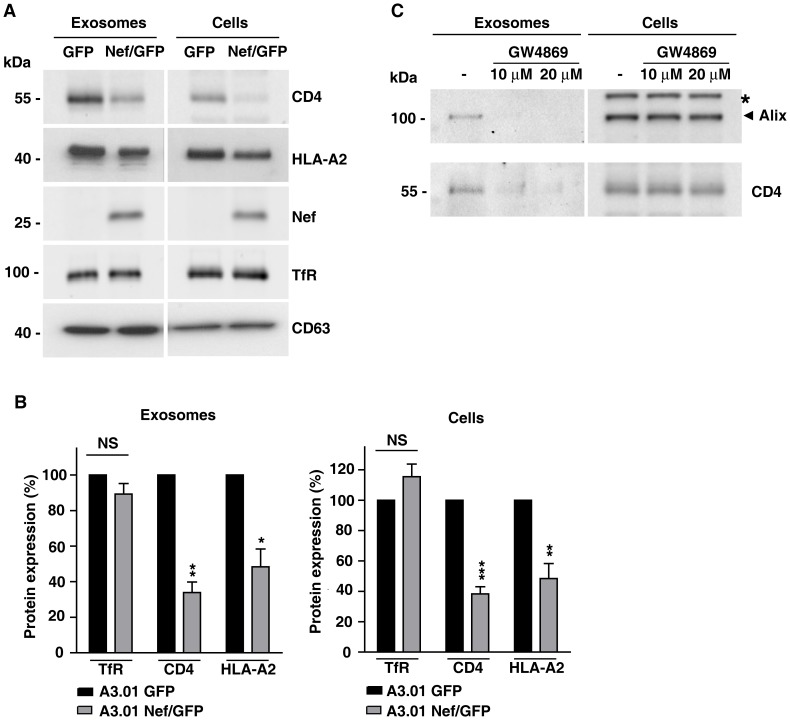
Figure 3. Nef reduces expression of CD4 and HLA-A2 in exosomes from CD4+ T cells.
(A) Exosomes released from equal numbers of A3.01 T cells expressing GFP or Nef/GFP were isolated from culture supernatant after 72 h of culture and equivalent amounts of exosome lysates or cell lysates were western blotted with antibodies to CD4, HLA-A2, Nef, TfR and CD63. (B) The TfR, CD4 and HLA-A2 signals for each condition shown in panel A was determined by densitometry and used to calculate the relative amount of these proteins in either exosomes (left panel) or total cell (right panel) lysates from Nef/GFP relative to GFP cells (100%). Bars represent the means ± standard deviations (n = 3) of normalized data. P-values calculated by Student's t-test using the raw data from densitometry analysis were as follows: * P<0.05; **, P<0.005, and ***, P<0.0005; NS, not significant. (C) Inhibition of CD4 secretion in exosomes by GW4869. A3.01 GFP cells were cultivated in absence or presence of 10 µM or 20 µM of GW4869 for 24 h. After treatment, conditioned medium were collected and exosomes were purified as described in material and methods. Exosomal and cellular proteins were analyzed by SDS-PAGE (6% gel) and western blot with antibodies to Alix or CD4. A nonspecific band detected with anti-Alix antibody is indicated with an asterisk. Note that treatment with GW4869 reduced the amount of both Alix and CD4 recovered in the exosome fractions. The results shown are representative of three independent experiments.

