FIGURE 1:
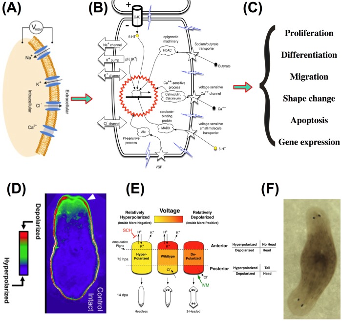
Bioelectrical signaling at the cell and organism levels, At the level of single cells, bioelectrical signals are produced by ion channel proteins, transduced into second-messenger responses, and alter key aspects of cell behavior. (A) The voltage potential (Vmem) at the cell membrane is produced by the movement of ions through across a cell membrane. Ions move via many different ion channels and pumps, under the control of concentration and electric gradients. (B) Change of Vmem is transduced into cellular effector cascades by a range of mechanisms, including voltage-sensitive phosphatases, voltage-gated calcium channels, and voltage-sensitive transporters of signaling molecules such as serotonin and butyrate. (Diagram modified, with permission, from Figure 1B of Levin, 2007.) (C) Bioelectrical signals feed into epigenetic and transcriptional cascades and thus trigger changes in cell properties such as proliferation, differentiation, migration, shape change, and programmed cell death. (D) Voltage reporter dye reveals gradients of Vmem across the anterior-posterior axis of planarian flatworms. (Taken, with permission, from Figure 2B of Beane et al., 2013.) (E) In amputated worms, a circuit composed of proton and potassium conductances sets the voltage states at each blastema, which in turn determines the anatomical identity of each end of a regenerating fragment. (Diagram taken, with permission, from Figure 7C of Beane et al., 2011.) (F) Manipulating this circuit in amputated planaria using pharmacological or genetic techniques that target ion flux allows the programming of stem cell–mediated morphogenesis to specific anatomical outcomes, such as the creation of two-head animals shown here.

