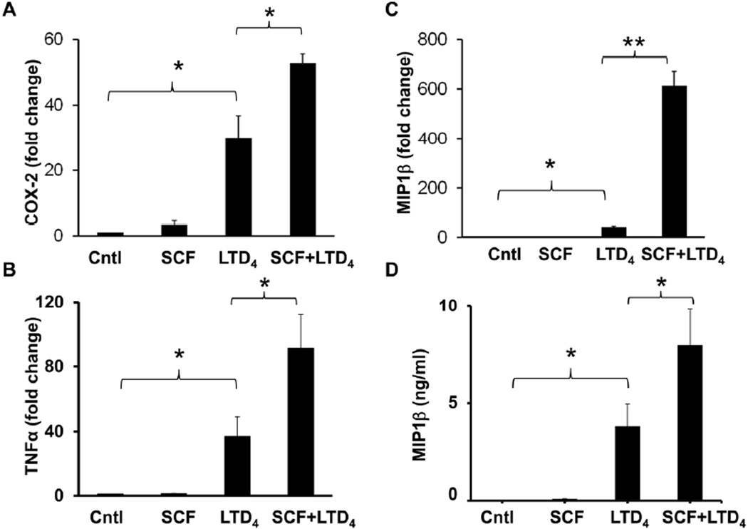
Figure 6. SCF augments LTD4-induced inflammatory gene repertoire.
LAD2 cells were treated with 500 nM LTD4 and/or 100ng/ml of SCF for 2 h, followed by mRNA extraction and cDNA synthesis. Transcript levels of COX-2 (A), TNFα (B), and MIP1β (C) were analyzed in these cDNAs using respective real time primers and were analyzed compared to GAPDH. The graph represents fold change in the level of transcripts compared to controls from three separate experiments. (D) LAD2 cells were treated with 500 nM LTD4 in the presence or absence of 100ng/ml SCF for 6h. Culture medium was collected and analyzed for secreted MIP1β protein by ELISA. The data shown represents mean ± SEM of three separate experiments. Data was analyzed with one way ANOVA and post-hoc analysis. *P<0.05, **P<0.001.

