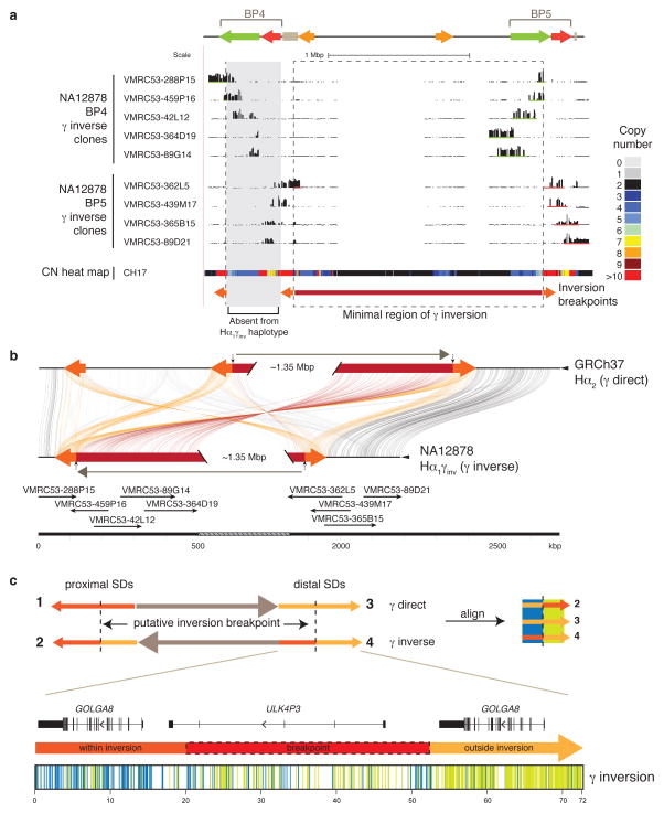
Figure 3. Sequence refinement of γ inversion breakpoints.
(a) The γ inversion was identified, validated, and sequenced using the VMRC53 BAC library (NA12878 individual). Illumina-generated sequences of clones spanning the 15q13.3 BP4 (green bars) and BP5 (red bars) loci were mapped to the human reference GRCh37. The nine clones pictured were sequenced using PacBio. The copy number (CN) heat map shows total diploid CN of a region in the CH17 hydatidiform mole cell line. The minimal region of the inversion spans ~1.8 Mbp (highlighted with a dashed box and a red bar). The orange arrows represent the flanking 72 kbp flanking inverted SDs that mediate the γ inversion. The Hα1γinv haplotype likely arose from the Hα1 haplotype, which does not harbor CNPα and CNPβ at BP4. (b) Homologous sequences of clones, generated using PacBio and assembled into contigs, and the human reference are connected with colored lines between γ direct (Hα2) and inverse (Hα1γinv) haplotypes using Miropeats55. Vertical arrows indicate the minimal inversion breakpoints. (c) Homologous sequences (72 kbp) from the orange flanking inverted SDs were aligned from multiple individuals (NA12878, CH17, and GRCh37) and haplotypes (γ direct: SD 3, and γ inverse: SDs 2 and 4; see Supplementary Figure 9 for a more detailed alignment) and variant sites compared. Variant positions showing signatures of being within or outside of the γ inversion breakpoints are indicated as colored lines under the picture of the distal γ inverse SD including: within the inversion (blue; consensus of SDs 2 & 3), and outside the inversion (green; consensus of SDs 3 & 4). The inversion breakpoint, refined to a region in which we observe a transition from blue to green lines, is highlighted with a dash-lined red box.

