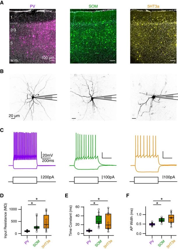
Figure 6.

Three populations of GABAergic interneurons. A, Coronal sections of prelimbic PFC with PV (left), SOM (middle), or 5HT3 (right) interneurons expressing ChR2-mCherry (false color) and stained with DAPI (gray), where layers and white matter (w.m.) are indicated on the left. B, Two-photon images of PV (left), SOM (middle), and 5HT3a (right) interneurons during whole-cell recordings. C, Positive and negative current injections indicate distinct passive and active properties at different interneurons. D–F, Summary of input resistance (D), membrane time constant (E), and AP width (F) for the different interneurons. *p < 0.025.
