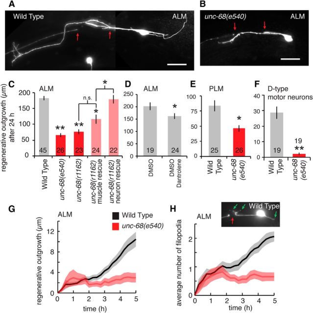
Figure 1.
Disruption of RyR channels reduces in vivo neuronal regeneration. A, B, Example images of C. elegans ALM neuron regeneration at 24 h for wild-type and unc-68(e540) mutant worms. Red arrows indicate position of laser damage. Scale bars, 20 μm. C, Average regenerative outgrowth 24 h after laser axotomy of ALM neurons for wild-type, unc-68 mutants and rescue strains. *p < 0.05 versus wild-type (except where indicated by the brackets). **p < 0.001 versus wild-type (except where indicated by the brackets). n.s., Not significant (multiple comparison in one-way ANOVA using a Tukey adjustment). D, Average 24 h regenerative outgrowth of axotomized ALM neurons for control (with DMSO) and dantrolene (10 μm)-treated C. elegans. *p < 0.05 (t test). E, F, Average 24 h regenerative outgrowth of axotomized PLM and D-type motor neurons for wild-type and unc-68(e540) mutants. *p < 0.05 (t test). **p < 0.001 (t test). G, Time-lapse measurement of average regenerative outgrowth for the 0–5 h following laser axotomy of ALM neurons. Measurements every 15 min. Shaded region represents SEM. H, The average number of filiopodial outgrowths in regenerating ALM neurons for wild-type and unc-68(e540) measured at every 15 min over the 0–5 h following laser axotomy. Inset, Example of wild-type regeneration at 5 h showing three filopodial outgrowths (green arrows).

