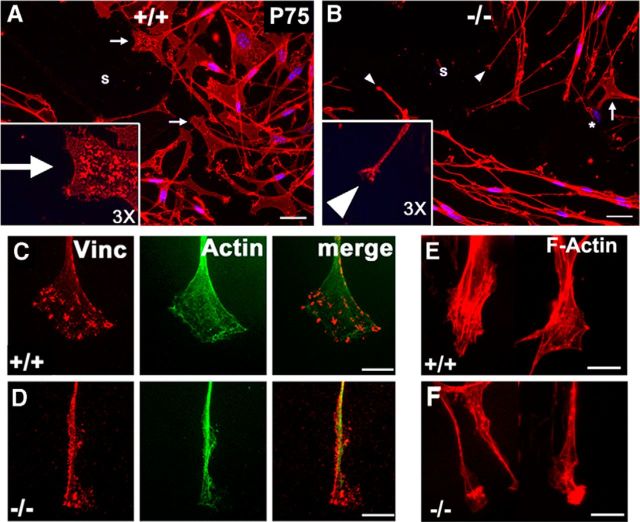
Figure 4.
Lack of PMP22 in Schwann cells is associated with morphological alterations in lamellipodial spreading. A, B, Schwann cells at the wound edge in WT (PMP22+/+) and PMP22−/− cultures after immunolabeling with anti-P75 antibody at 8 h postscratch. s, scratch area; arrows, lamellipodia; arrowheads, filopodia. Insets in A and B show a 3× enlarged view of representative lamellipodia. C, D, Images of lamellipodia of WT and PMP22−/− Schwann cells after colabeling with anti-vinculin and anti-actin antibodies. E, F, Micrographs of WT and PMP22−/− Schwann cell processes after labeling with phalloidin. Images are representative of cells from three to four independent cultures. Scale bars: A, B, 20 μm; C–F, 10 μm.

