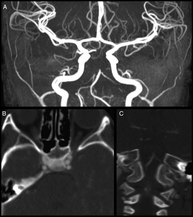
Figure 2.
(A) MR angiography shows an abrupt cut-off of the mid portion of the basilar artery, with lack of flow-related enhancement throughout the remainder of the expected course of the basilar artery. There is a large right posterior communicating artery which supplies the right P2 distribution. A very small left posterior communicating artery (Pcomm) is seen only over approximately a 7–8 mm segment. It appears that the right P1 segment is filling from the right Pcomm. (B) Axial CT angiography shows no filling of the basilar artery in the pontine cistern. (C) Coronal CT angiography shows complete occlusion of the basilar artery just distal to the origins of the anterior inferior cerebellar arteries.

