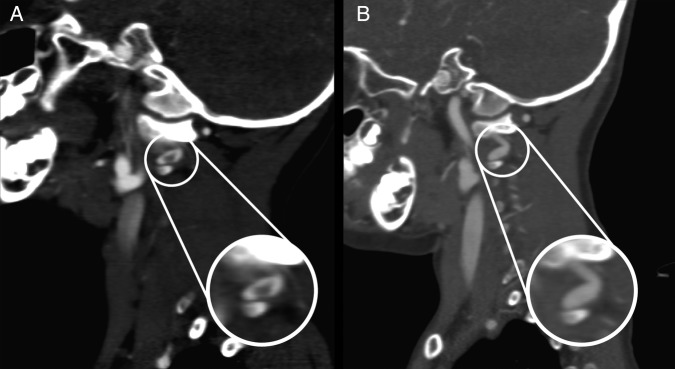
Figure 3.

(A) Sagittal CT angiography (CTA) demonstrates possible dissection at the V3 segment of the vertebral artery. (B) Two-week follow-up sagittal CTA demonstrates an apparent smooth caliber change over a short segment of the left vertebral artery at C1–C2 at the area of previous thrombus on prior CTA. This possibly represents a pseudoaneurysm.
