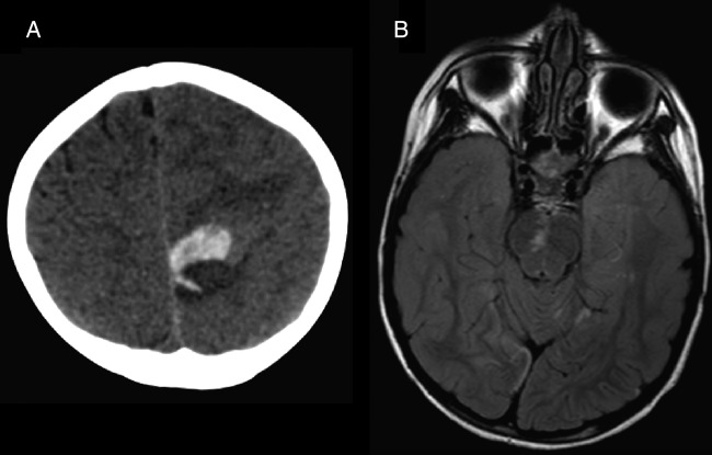
Figure 5.
(A) Head CT 2 days post-thrombectomy shows new acute high medial left frontal lobe hematoma with mild/moderate surrounding edema and local mass effect. This was thought to represent hemorrhagic transformation of distal emboli that spontaneously recanalized as it was not in an area previously at risk. (B) MRI FLAIR sequence obtained 3 days after mechanical thrombectomy shows some persistent changes, although there has been some reversal of diffusion-weighted imaging changes compared with the admission MRI, especially within the brainstem (compare figure 1, middle bottom row).

