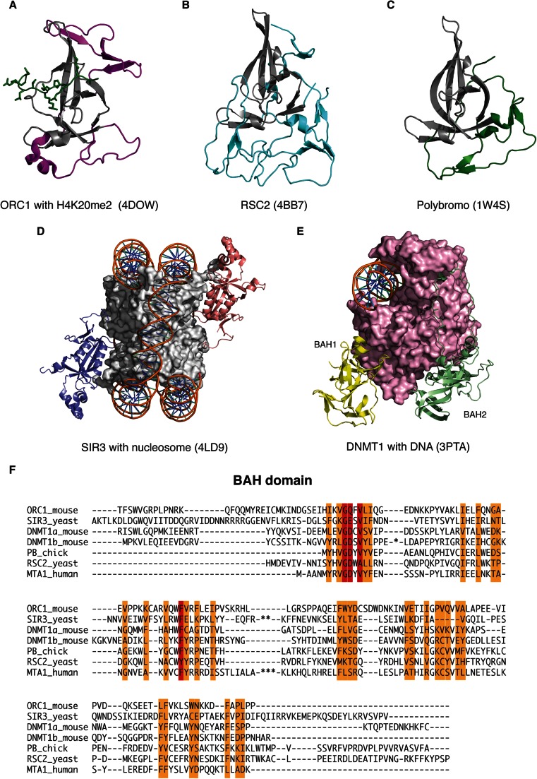
Fig. 5.
The structure of the BAH domain. a The BAH domain from ORC1 (magenta) with bound methylated H4 peptide (green). b The BAH domain from RSC2 (cyan). c The BAH domain Polybromo (green). The canonical core BAH domain is coloured grey in each case. d Two copies of SIR3-BAH (purple and salmon) in complex with a nucleosome. DNA is shown as a cartoon, and the four histones are shown as surface (grey). e DNMT1 bound to DNA. The BAH1 and BAH2 domains are highlighted (yellow and light green), and the rest of the protein is shown as surface (pink). f Alignment of the MTA1-BAH with the sequences of BAH domains for which their structure is known. Forty residues in DNMT1b (*), 15 residues in SIR3 (**) and 38 residues in MTA1 are not shown (***) (colour figure online)

