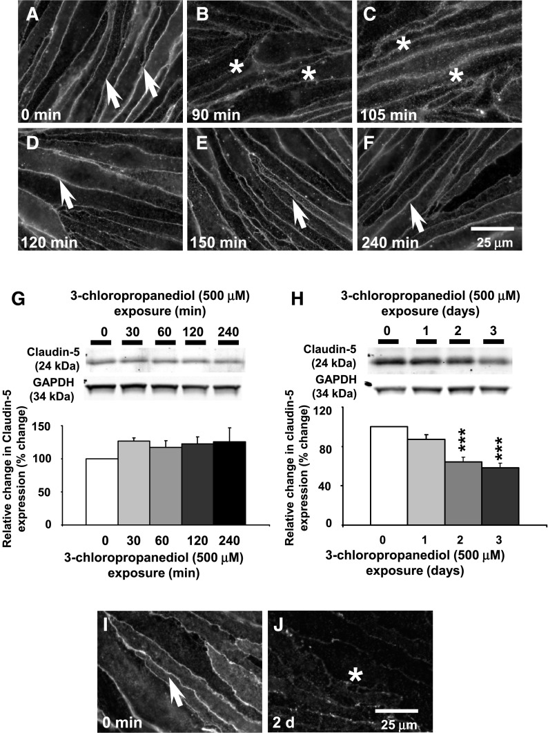
Fig. 1.
Early and late loss of paracellular claudin-5 expression. (A–F) Immunofluorescence analysis showed early reversible loss of paracellular claudin-5 expression over a 240-minute period. Arrows and asterisks show claudin-5 immunoreactivity. (G) Western blot analysis showed no loss of total claudin-5 expression over a 240-minute time course. (H) A marked loss (65%) of claudin-5 expression was observed after 3 days. (I and J) Immunofluorescence studies showed a late loss of claudin-5 expression after 3-chloropropanediol exposure for 2 days. Protein quantification data were obtained by densitometry and normalized using GAPDH as a loading control. Values are expressed as relative optical density and are represented as mean ± S.E.M. ***P < 0.001 (compared with the controls). For each column, n = 4–6 independent experiments. GAPDH, glyceraldehyde 3-phosphate dehydrogenase. Scale bar, 25 μm in A–F, I, and J.

