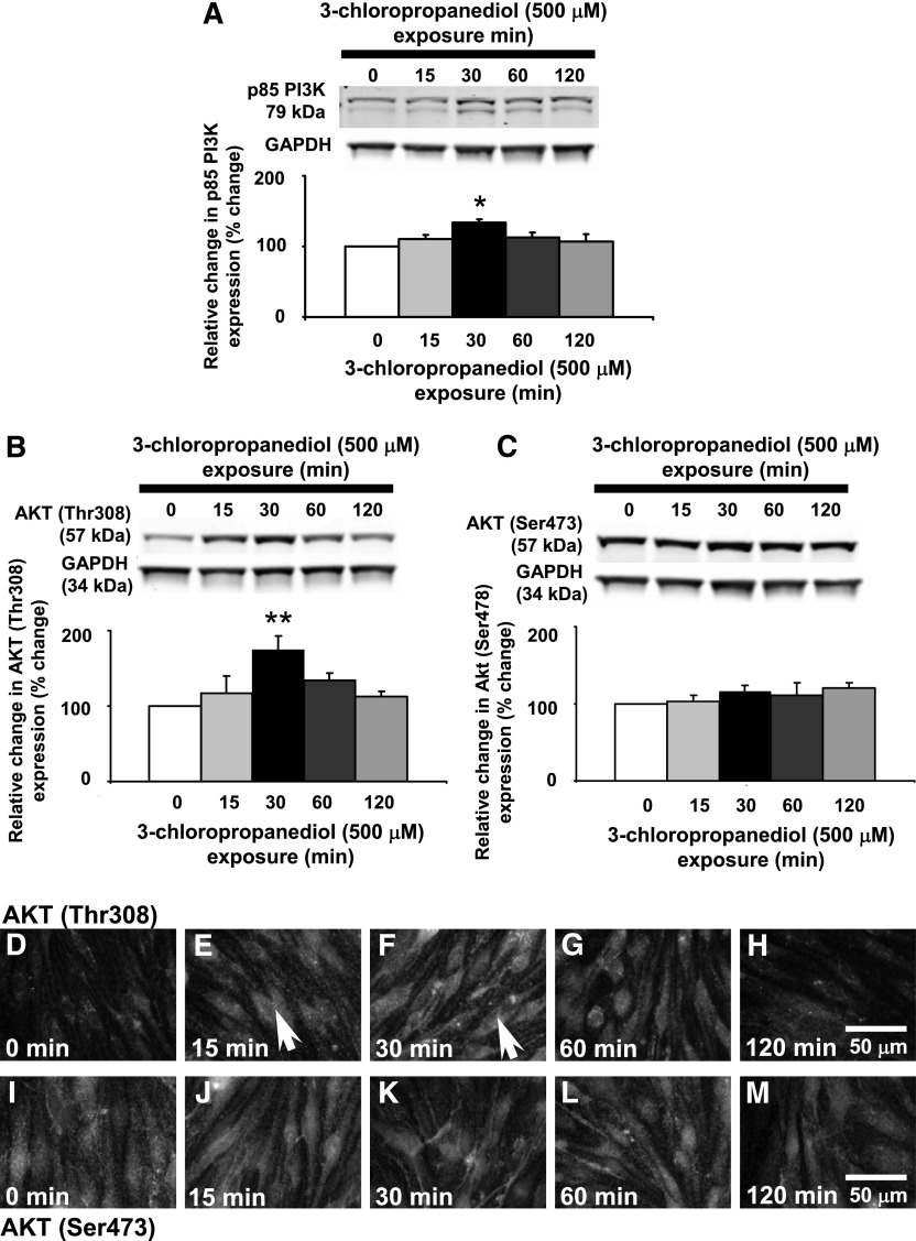
Fig. 2.
Transitory increased p85-PI3K and AKT (Thr308) phosphorylation. Western blot analysis showed changes in p85-PI3K and AKT (Thr308) expression in bEnd.3 cells after 3-chloropropanediol (500 μM) administration. (A) p85-PI3K appeared as a 79-kDa band and showed increased expression at 30 minutes. (B) AKT (Thr308) appeared as a 57-kDa band. Over 30 minutes, there was a marked increase in expression of the 57-kDa band. The intensity of this band decreased after 60 and 120 minutes. (C) AKT (Ser473) showed no change in expression. (D–H) Immunofluorescence analysis showed increased AKT (Thr308) expression at 15 and 30 minutes after 3-chloropropanediol administration compared with controls. By 60 and 120 minutes, expression decreased and was comparable to control cells. Arrows show increased cytoplasmic immunofluorescence. (I–M) AKT (Ser473) phosphorylation showed no change in expression over the study period. Protein quantification data were obtained by densitometry and normalized using GAPDH as a loading control. Values are expressed as relative optical density and are represented as mean ± S.E.M. *P < 0.05; **P < 0.01 (compared with the controls). For each column, n = 4–6 independent experiments. GAPDH, glyceraldehyde 3-phosphate dehydrogenase. Scale bar, 50 μm in D–M.

