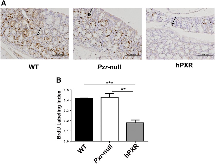
Fig. 6.
Colon epithelial cells exhibited lower proliferation in PXR-humanized (hPXR) mice than wild-type (WT) and Pxr-null mice upon short-term treatment with rifaximin and AOM/DSS. (A) Representative BrdU staining in colon tissues obtained from hPXR, wild-type, and Pxr-null mice treated with rifaximin and AOM/DSS. Arrows indicate BrdU-positive nuclei. (B) BrdU labeling index comparison in hPXR, WT, Pxr-null mice treated with rifaximin and AOM/DSS. **P < 0.01; ***P < 0.001.

