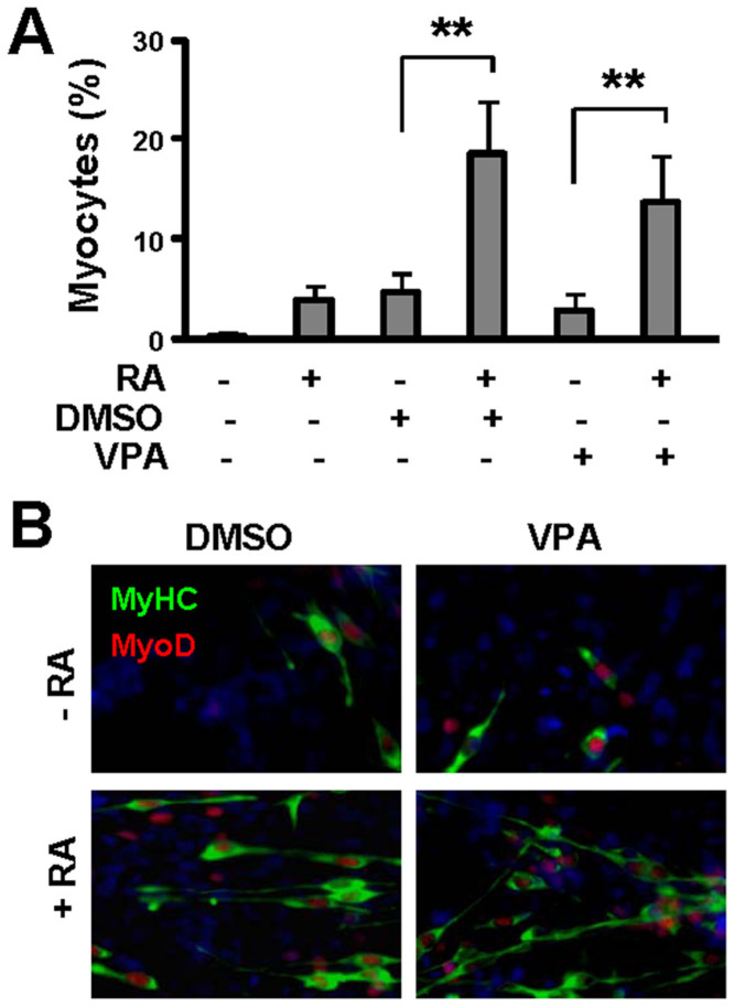
Figure 1. Effects of valproic acid on myogenic differentiation.
(A) Pluripotent P19 cells were grown as EBs for 4 days and treated with DMSO (1%), RA (10 nM) or valproic acid (VPA, 0.5 mM). The cells were cultured for an additional 5 days without treatment and stained for myosin heavy chain and nuclei on day 9 of differentiation before microscopic analysis. Quantification is presented as the percentage of cells differentiated into skeletal myocytes. Error bars are the standard deviations of four independent experiments. Statistical significance is denoted by ** (p< 0.01). (B) Representative microscopic images of myosin heavy chain (MyHC, green), MyoD (red) and nuclei (blue) co-staining.

