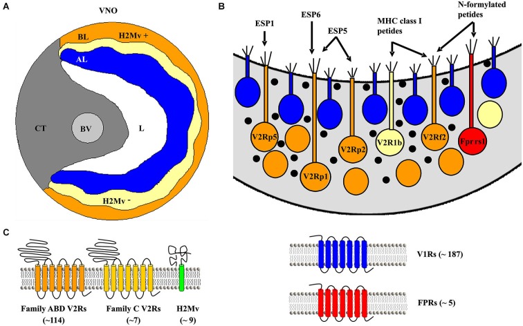
Figure 1.
The mouse vomeronasal organ (VNO) and established receptors located in the epithelium. (A) Schematic coronal section of the VNO. AL, apical layer of the sensory epithelium (blue); BL, basal layer (yellow/orange); BV, blood vessel; CT, cavernous tissue; H2Mv+, sensory epithelium cells lacking one of the nine known H2-Mv genes; H2Mv−, sensory neurons not expressing any of the nine H2-Mv genes; L, lumen. (B) Schematic drawing of sensory neurons showing their location in the basal sensory epithelium with their proposed ligands. (C) Schematic picture of the proposed receptors expressed in the basal part of the VNO sensory epithelium.

