
Figure 4.
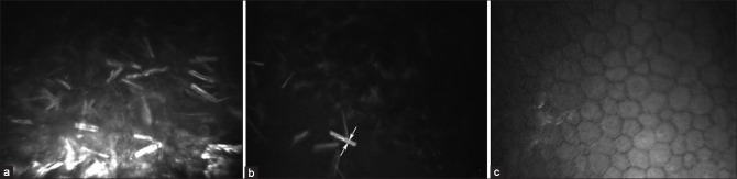
In vivo confocal microscopy images of case number 2. (a) Dense infiltration of the corneal epithelium with typical centrally hypointense slender crystalline deposits. (b) An oblique section at the epithelium-cornea junction depicting the localization of the immunoprotein deposits to the epithelial layer with uninvolved stromal appearance. The characteristic morphology of the immunoprotein deposit with a central hypointense lumen and hyperintense borders is depicted with arrows. (c) The endothelium appeared to be devoid of crystalline infiltration and of normal cellular morphology