
Figure 2.
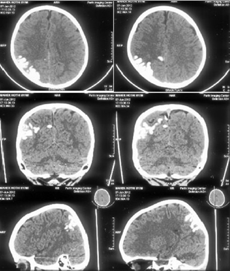
Axial, coronal, and sagittal images of a plain CT head show gyriform calcification in the right parietal cortex. Associated volume loss in the parietal lobe is noted
Official websites use .gov
A
.gov website belongs to an official
government organization in the United States.
Secure .gov websites use HTTPS
A lock (
) or https:// means you've safely
connected to the .gov website. Share sensitive
information only on official, secure websites.
Axial, coronal, and sagittal images of a plain CT head show gyriform calcification in the right parietal cortex. Associated volume loss in the parietal lobe is noted