
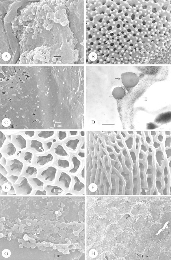
Fig. 1. SEM of species with Type I orbicules (A and B) and SEM and TEM of Type IIIa (C–H) orbicules. Abbreviations used in figures: E, endothecium; ED, electron dense; ET, electron translucent; PE, pollen exine; SEM, scanning electron microscopy; TEM, transmission electron microscopy. A and B, Hockinia montana. A, SEM observation of the spiny orbicules (arrow), covering the locule wall surface. B, Detail of the exine ornamentation. Microspines (arrow) are present upon the tectum. C–E, Gentianopsis procera. C, SEM observation of the small spherical orbicules (arrow). D, TEM observation of the orbicules. An electron‐transparent orbicule cavity is absent. The orbicule wall is delimited by a very electron‐dense and granular peripheral layer (arrow). Bar = 0·6 µm. E, Detail of the reticulate exine. The lumina are beset with granules (white arrow). The muri are sharply crested (black arrow). F, Gentianella bellidifolia. SEM observation of the striato‐reticulate exine. The muri are oriented parallel and criss‐crossed with smaller muri. The muri are rounded (arrow). G and H, Ixanthus viscosus. G, SEM observation of single or compound orbicules. Sporopollinous threads occur between the orbicules (arrow). H, General view of the locule wall surface, imprints of pollen grains are striking. Orbicules (black arrow) are present on the edges between the imprints.