
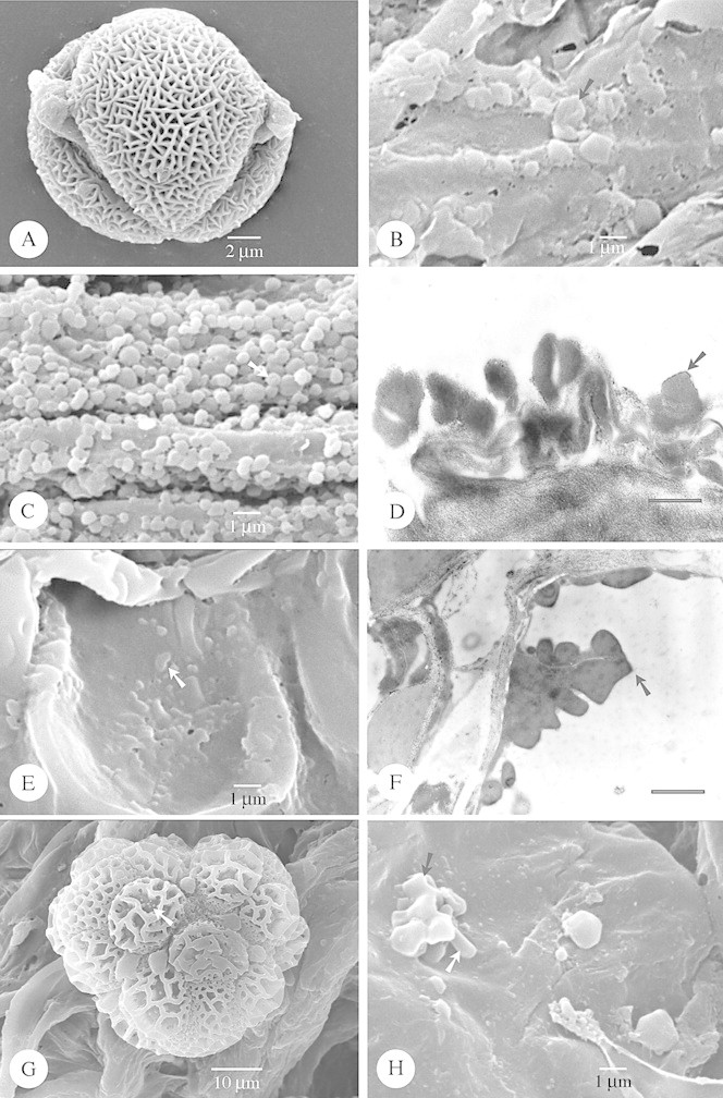
Fig. 4. SEM and TEM of species with irregular and folded Type IV orbicules. A and B, Sebaea ovata. A, Equatorial view of a striato‐micro‐reticulate pollen grain. B, Detail of the irregular and folded orbicules (arrow), resembling parts of the muri building up the exine. C and D, Sebaea albidiflora. C, SEM observation of the irregular and folded orbicules (arrow). D, TEM observation of the irregular orbicules positioned on the remnants of the tapetum cells. The orbicules posses an elongated electron‐transparent cavity and the homogeneous ED orbicule wall is delimited by a electron‐dense peripheral layer (arrow). Bar = 0·6 µm. E and F, Triptospermum fasciculatum. E, SEM observation of the irregular and folded orbicules (arrow) covering the locule wall surface. F, TEM observation of the irregular orbicules, lacking an electron‐transparent cavity. The homogeneous ED orbicule wall is delimited by an ED peripheral layer (arrow). Bar = 1 µm. G and H, Calolisianthus pedunculatus. G, SEM observation of a tetrad lying on the locule wall surface. The muri of the reticulate pattern are organized in heterobrochate reticulate islands. The lumina are beset with granules (arrow). H, SEM observation of the orbicules on the locule wall surface. The orbicules consist of columellae‐like particles (white arrow) which support the muri‐like part (black arrow) of the orbicules.