
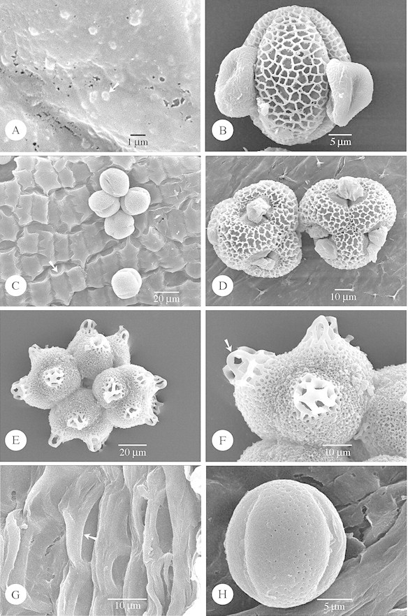
Fig. 6. SEM of species with strongly embedded Type VI (A and B) orbicules, and species lacking orbicules (C–H). A and B, Lisianthius nigrescens. A, General observation of the strongly embedded orbicules (arrow). B, Equatorial view of a reticulate pollen grain. C, Lomatogonium carinthiacum. General view of five pollen grains lying on the locule wall surface, which lacks orbicules. Deep grooves in the locule surface reveal the position of the underlying endothecial cells, the U‐shaped endothecial thickenings (arrow) can be distinguished. D, Schultesia guianensis. SEM observation of two reticulate tetrads lying on the smooth locule wall surface. E and F, Irlbachia purpurascens. E, General view of a micro‐reticulate polyad. F, Detailed observation of the muri of the reticulate pattern, at the distal regions, which are loop‐like. The loops of reticulum (arrow) are raised from the general surface of the pollen grain. G and H, Curtia tenuifolia. G, SEM observation of the locule surface consisting of wavy ridges (arrow). H, Equatorial view of a perforate pollen grain lying on the locule wall surface.