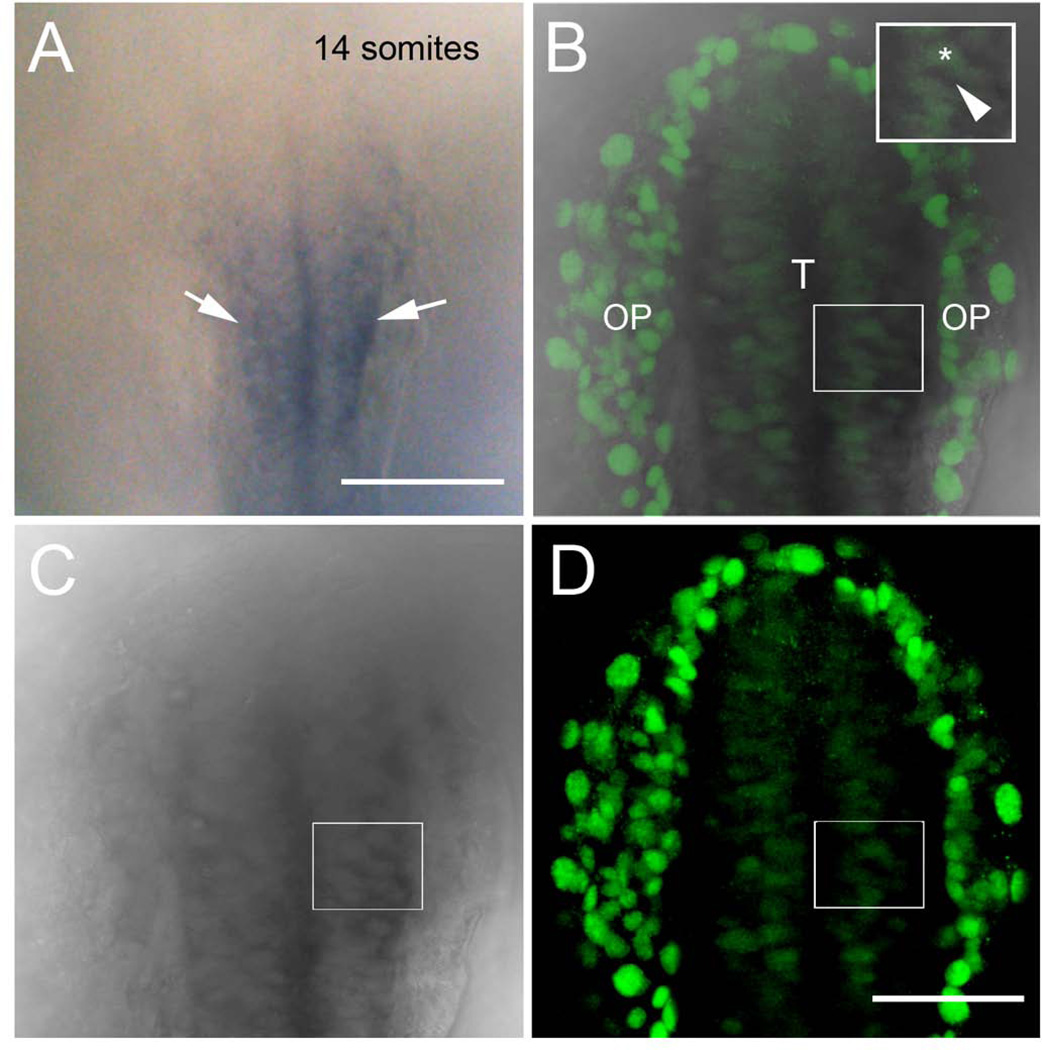
Figure 2.
Expression of emx1 co-localizes with Dlx3b in the developing telencephalon. A) Transmitted light image of wholemount embryo processed for emx1 expression marking the telencephalon (arrowheads). B–D) Different preparation of 14ss wholemount embryo with Dlx3b immunodetection (B, D green) after in situ hybridization for emx1 (B,C, dark gray). Merged image of 3 µm focal plane (B) colocalizes Dlx3b signal (green) with transmitted light image of emx1 in situ (grey). Inset box in B shows higher magification of cells expressing Dlx3b (asterisk) and emx1 (arrowhead). All embryos are dorsal views with anterior toward top of page. OP, olfactory placodes; T, telencephalon. Scale bars: A, 100 µm; B–D, 50 µm.

