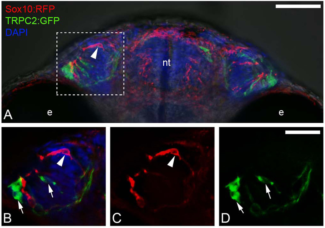
Figure 9.
Sox10:RFP positive neural crest does not contribute to TRPC2:GFP positive microvillar neuronal populations within the olfactory sensory epithelium. A) wholemount head at 55 hpf viewed from ventral. Boxed area indicates region of interest shown in B–D. B) Expression of Sox10:RFP (red, arrowhead) and TRPC2:GFP (green, arrows) in the OP. B) TRPC2:GFP (arrows) in the olfactory organ. C) Expression of Sox10:RFP (arrowhead) in the olfactory organ. B–D) Images are 3 µm optical sections. Scale Bars: A=50µm; B–D =25µm.

