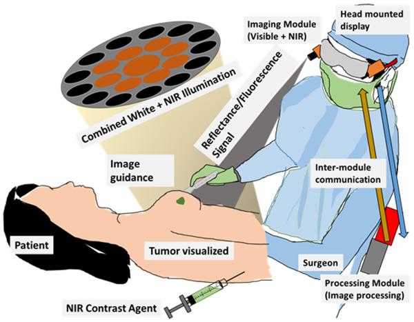
Figure 5.
Goggle system overview. After injection of an imaging agent, our system excites the contrast agent. The NIR fluorescence and color reflectance images are captured and processed to generate a superimposed image where fluorescence is highlighted in a false color on the normal view. This superimposed image is seen in the head mounted display in real time by the surgeon, who can clearly visualize the tumor boundary during cancer surgery.

