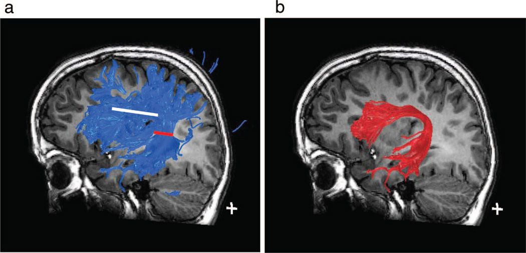
Figure 4.
Fiber tracking. (A) Three-dimensional visualization of superior longitudinal fasciculus fibers, tracked from the region of interest outlined in white in Figure 2D. The location of the "seed" region of interest is depicted in this sagittal view as a white line. The blue fiber tract contains fibers connecting the temporal, parietal, and frontal lobes. (B) The arcuate fasciculus, the portion of the superior longitudinal fasciculus that connects the temporal lobe to the frontal lobe. Tract was defined by limiting the blue fiber group in Figure 4A to only those fibers passing through a second region of interest, depicted by the red line, thereby using a 2 region of interest approach.

