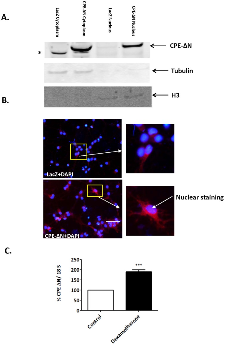
Figure 2. Subcellular localization and up-regulation of CPE-ΔN mRNA expression by dexamethasone.
A. Cellular localization of CPE-ΔN in rat E18 cortical neurons transduced with adenovirus carrying CPE-ΔN construct. Western blot shows the presence of CPE-ΔN in the cytoplasm and the nucleus of CPE-ΔN-transduced cells, but not visible in the LacZ-transduced control cells. Beta-tubulin served as a marker for cytoplasm and Histone-3 served as a marker for nucleus. * indicate a non specific band in the cytoplasm. B. Immunocytochemistry confirm CPE-ΔN (red) immunostaining in the nucleus marked by DAPI staining (blue) and is more visible in the CPE-ΔN-transduced vs. control cells. Insets show a higher magnification of cells indicated by the arrow. Bar = 50 microns. C. Bar graphs show an increase in CPE-ΔN mRNA expression in rat E18 hippocampal neurons treated with dexamethasone compared to control untreated neurons. Values are the mean ±SEM (n = 4). t test *** p<0.001.

