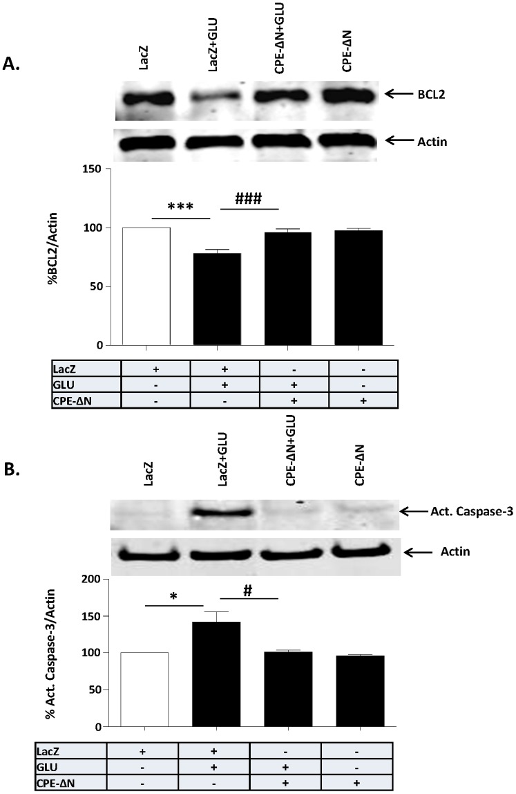
Figure 6. Neuroprotection by CPE-ΔN involves BCL-2 and Caspase-3.
In A, B, rat primary cortical neurons were transduced with CPE-ΔN or LacZ viral vectors and subsequently treated with or without with glutamate for 24 h. A. Top panel: Western blot of BCL-2 protein in primary cortical neurons challenged or not with glutamate. Actin was also analyzed and served as an internal control for protein load; bottom panel: Bar graphs showing the quantification of BCL-2 normalized to actin and expressed as a % compared to vehicle treated control cells. Note that CPE-ΔN significantly inhibited the glutamate-induced decrease in BCL-2 protein in the cortical neurons. At least three independent experiments were done. Data shown represent all the experiments combined. B. Top panel: Western blot of caspase 3 protein in primary cortical neurons challenged or not with glutamate. Actin was also analyzed and served as an internal control for protein load; Bottom panel: Bar graphs showing the quantification of active caspase-3 normalized to actin and expressed as a % compared to vehicle treated control cells. Note that CPE-ΔN significantly inhibited the glutamate-induced activation of caspase-3 in the primary cortical neurons. At least three independent experiments were done. Data shown represent all the experiments combined. n = 6/group (A); 3/group (B). Values are mean ± SEM, one-way ANOVA followed by Tukey test, *p<0.05.

