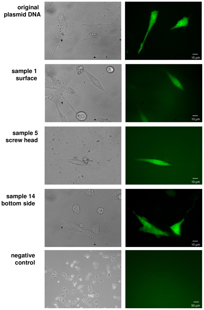
Figure 6. DNA function in eukaryotic cells.
For each application location (surface, screw head, bottom side) one recovered DNA sample was chosen for transfection into NIH-3T3 mouse fibroblasts and EGFP expression was visualized after 1 day of incubation. In all the samples EGFP expression was detectable indicating the functionality of the DNA samples. Left column: Phase contrast images; Right column: GFP filter.

