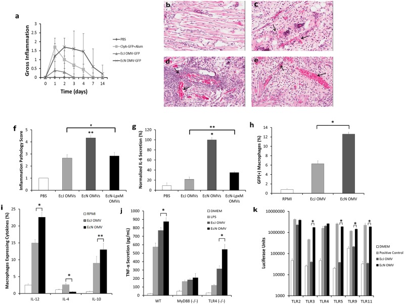
Figure 3. EcN OMV carriers are capable of potent self-adjuvanting engagement of innate immunity.
a, Observed macroscopic inflammation (scored 0–3 for severity) at s.c. injection site (n = 5, each group). B–e, Representative dermal histological sections from the injection site (t = 30 h) in BALB/c mice (n = 4, each group), injected with PBS (b), EcJ OMVs (c), EcN OMVs (d), or lpxM-mutant EcN OMVs (e). Arrows indicate vasculature swelling and leukocyte recruitment, which is substantially enhanced by EcN OMVs and reduced, but not eliminated, by the lpxM mutation. f, Inflammation pathology scoring of b–e on a 0–5 scale. g, IL-6 secretion levels, normalized to the highest value in the data set, from primary mouse bone-marrow derived macrophages incubated with OMVs used in b-e. h,i, Flow cytometry (h) and cytokine secretion analysis (i) of primary mouse bone-marrow derived macrophages incubated with EcJ and EcN OMVs (n = 3, each group), demonstrating enhanced macrophage stimulation by EcN OMVs. j, TNF-α dependent activation of wild-type (WT), MyD88−/−, and TLR4−/− mouse macrophages incubated with EcJ and EcN OMVs (n = 5, each group) reveal MyD88-dependent, TLR4-dependent and TLR4-independent stimulation via EcN OMVs. k, TLR activation in single-TLR expressing human embryonic kidney (HEK) cells incubated with EcJ and EcN OMVs (n = 3, each group), demonstrating broad TLR activation by EcN OMVs relative to exclusively TLR2/4 activation by EcJ OMVs. *P<0.001, **P<0.05, #No statistically significant difference. All values are given as mean + or +/− SD.

