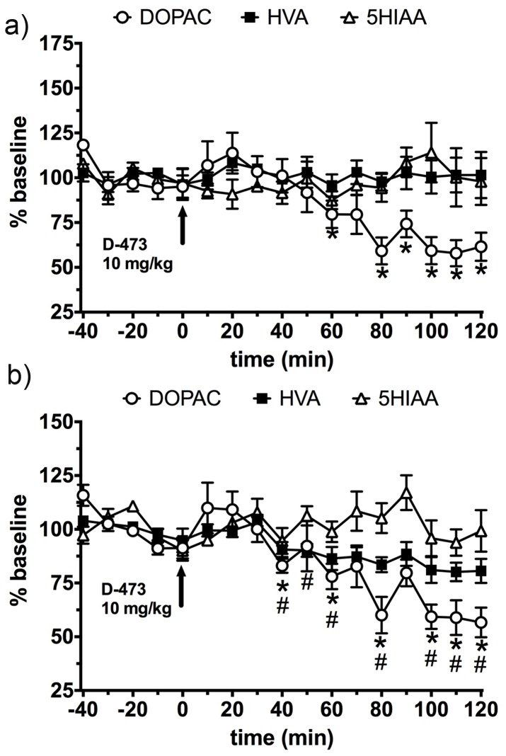
Figure 8. Time dependent effect of administration of D-473 (10 mg/kg, i.p) at time 0 (shown by arrow) on extracellular level of DOPAC (Ο), HVA (▪), and 5HIAA (Δ); a) in rats Prefrontal cortex; and b) Dorso lateral striatum.
Results are expressed as percent baseline with baseline values all averaging 100%. Each point represents mean ± standard error (SE) of the percentage of baseline from five rats. Statistical analysis was performed by t-test analysis of every point relative to baseline values (i.e. 100%) using Prism 6 (Graphpad Software Inc, La Jolla, CA). * (DOPAC) p<0.02-0.0004. # (HVA) p<0.02–0.001. P values <0.05 were considered to be statistically significant.

