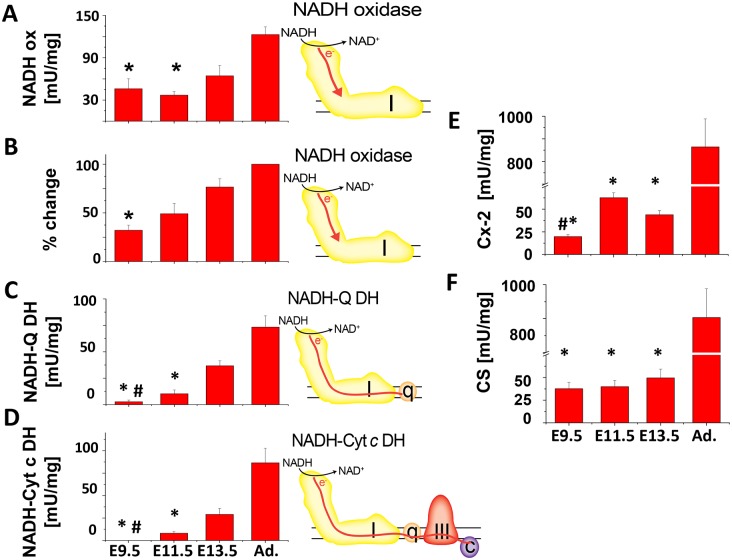
Figure 6. Enzymatic activity of Cx-1, Cx-2 and citrate synthase in cardiac tissue homogenates.
Each assay was done with 1–5 µg protein of cardiac tissue homogenate. The activities are given in mU/mg, except for the dipstick assay, where the relative change was calculated from signal of adult homogenates (100%). In A–D, the cartoon accompanying each graph depicts the flow of electrons through Cx-1 (I) and -3 (III), ubiquinone (q), and cytochrome c (c) tested in the assay. A. NADH-oxidase (NADH ox) assay, *p≤0.05 E9.5 or E11.5 versus adult. B. Cx-1, NADH-oxidase (NADH ox) dipstick assay, *p≤0.05 E9.5 versus adult. C. NADH-ubiquinone dehydrogenase (NADH-Q-DH) assay, *p≤0.05, E 9.5 or E11.5 compared to older embryos or adults; #p≤0.05, E9.5 versus E11.5. D. NADH-cytochrome c dehydrogenase (NADH-Cyt c DH) assay, *p≤0.05, E 9.5 or E11.5 compared to older embryos or adults; #p≤0.05, E9.5 versus E11.5 E. Cx-2/succinate dehydrogenase assay, *p≤0.05, embryos compared to adults; #p≤0.05, E9.5 versus E11.5 and F. Citrate synthase assay. *p≤0.05, embryos compared to adults; #p≤0.05, E9.5 versus E11.5. In all experiments, ANOVA with Tukey post-hoc testing was used and n≥3. All other comparisons were not significant.

