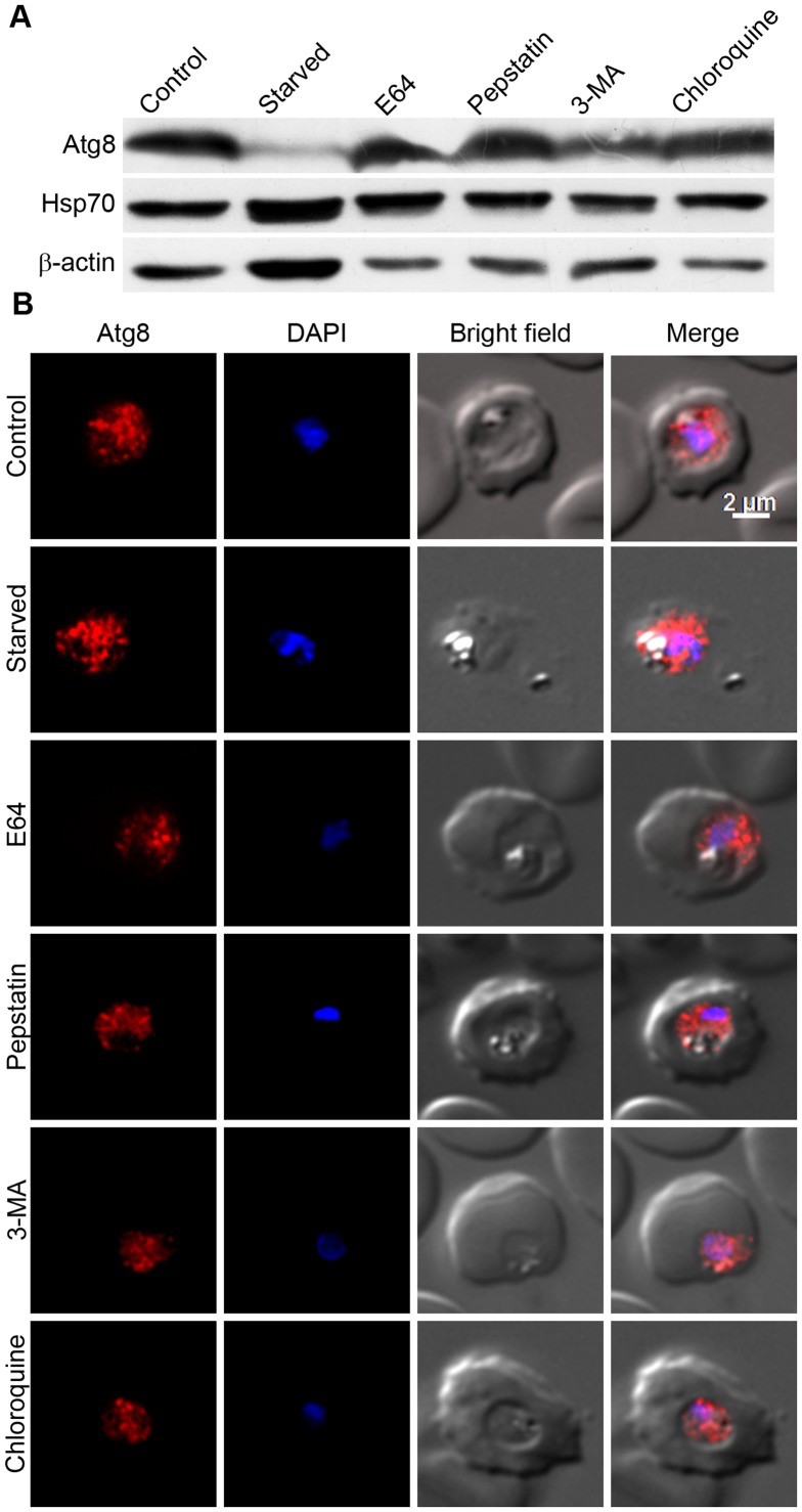
Figure 8. Effects of typically used autophagy inducer/inhibitors on Atg8 levels and localization.
Early/mid trophozoite stage parasites were cultured in HBSS (Starved) or in complete medium containing DMSO (Control) or the indicated autophagy inhibitors (5 mM 3MA, 30 nM CQ, 22 µM E64, and 220 µM pepstatin (Pep); all except 3MA are at concentrations 3× IC50) for 8 hours, and then parasites were evaluated for expression of Atg8 by western blotting (A) or for localization of Atg8 by IFA (B) using anti-Atg8 antibodies as described in the Materials and Methods section. The same parasite samples were assessed for expression of the control proteins β-actin (β-Actin) and Hsp70 by western blotting as described in the Materials and Methods section. A. The immunoblot shows drastically reduced Atg8 levels in starved parasites and almost similar Atg8 levels in other parasite samples compared to control parasites. Both Hsp70 and β-actin levels are similar in all except the starved parasites, which may be due to a starvation-induced stress response. B. The parasite images are labelled as in Figure 2, and show similar Atg8 signal regardless of the treatment. The experiment was repeated three times, and the results were reproducible.

