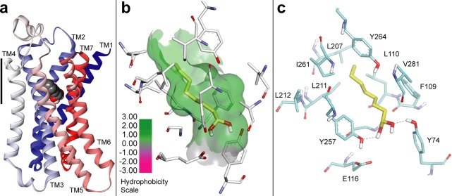
Figure 4.

Homology model of rat OR-I7 based on the activated ß2-adrenergic receptor (pdb 3SN6) and bound to octane-1,1-diol. (a) Overall structure showing OR-I7 with the octane-1,1-diol agonist aiming the gem-diol toward trans-membrane helices (TM) 2 and 7. TMs are colored from blue (N-terminus) to red (C-terminus). Ligand membrane depth is shown in relation to TM4 (scale bar, 12.7 Å). (b) The octanal carbon chain is in a hydrophobic pocket formed by TMs 3, 5, and 6. (c) Possible H-bond recognition of the gem-diol by Y74 and Y257. Carbons of octane-1,1-diol are shown in yellow.
