
Figure 2. Representative angiographic images of non-ISR, focal and diffuse ISR.
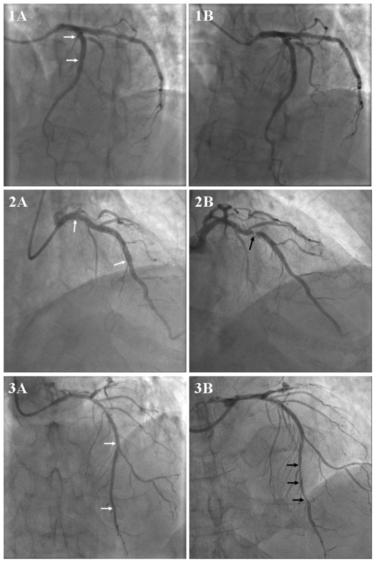
Angiographic results after successful percutaneous coronary intervention with stent implantation in the left anterior descending coronary artery from three different patients (1A, 2A and 3A). Follow-up angiography showed non-ISR (1B), focal ISR (2B) and diffuse ISR (3B) respectively. White arrows indicate the margins of stent. Black arrows indicate the stenotic lesions.