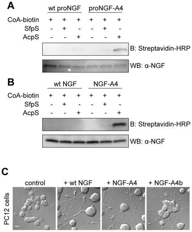
Figure 2. Site-specific biotinylation of proNGF and NGF.

A–B) Western blot for the analysis of the in vitro biotinylation reaction of purified NGF-A4 (A) and proNGF-A4 (B) using CoA-biotin substrate and AcpS or SfpS PPTases. The same biotinylation reaction is performed in parallel using untagged wt NGF and wt proNGF as negative controls. Streptavidin-HRP is used for detection of biotin. The anti-NGF blot is the loading control. C) Typical DIC images obtained when performing the differentiation assay in PC12 cells using ∼50 ng/ml of wt NGF, NGF-A4 and biotinylated NGF-A4 (NGF-A4b). Untreated cells are the control. Scale bar: 20 µm.
