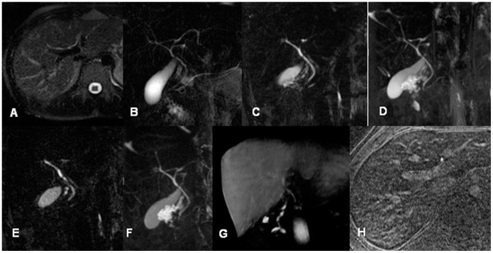
Figure 1. Comparison of MRC sequences in a 26-year-old male potential living liver donor (A: axial T2w HASTE; B coronal thick slab HASTE; C: central single image of coronal T2w 3D RESTORE; D: MIP of C; E: central single image of coronal T2w 3D SPACE; F: MIP of E; G: Gd-EOB-DTPA enhanced T1w FLASH sequence (coronal MPR) and H: axial Gd-EOB-DTPA enhanced T1w FLASH with IR pulse).
Note that all MRC data show abnormal central anatomy with Smadja & Blumgart Type D1 trifurcation while 3D sequences and coronal HASTE provide insight in the more peripheral bile ducts with drainage of the right posterior segment 7 duct into the left main hepatic duct.

