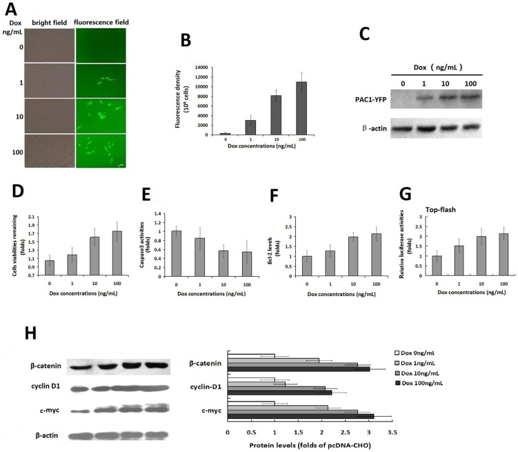
Figure 5. The correlation of the PAC1 levels with its basal activity in Tet-on inducible expression system.
The fluorescence microscopic observation (A) and the fluorescence density assays (B) of the PAC1-YFP expression induced by Dox (0–100 ng/mL) in Tet-on inducible system. The fluorescence microscopic images showed that the numbers of cells with YFP fluorescence increased following increases in the concentration of Dox (1–100 ng/mL), whereas there was no fluorescence observed without induction by Dox (0 ng/mL). Bar, 20 µm. The YFP fluorescence densities, assayed using the Victor3 1420 multi-label counter, increased with the concentration of Dox (1–100 ng/mL), indicating that the expression levels of PAC1 were controlled by Dox in a concentration-dependent manner. (C) Western blotting of the inducible expression of PAC1-YFP. The western blotting with goat polyclonal IgG against the C-terminus of PAC1 using reductive SDS-PAGE showed the bands corresponding to PAC1-YFP deepened with the increase of Dox (1–100 ng/mL), while no band corresponding to PAC1-YFP was found in the treatment without Dox (0 ng/mL). The remaining cell viabilities (D), the caspase3 activity (E) and the Bcl-2 levels (F) after serum withdrawal in the double-stable Tet-on advanced inducible cells treated with Dox (1–100 ng/mL) were plotted as the fold changes in the data from the cells treated without Dox (0 ng/mL). It was shown that the higher concentrations of Dox induced higher expression levels of PAC1-YFP, which in turn led to the higher anti-apoptotic activity of the cells, including higher remaining cell viability, lower caspase3 activity and higher Bcl-2 level. (G) Top-flash assays. In double-stable Tet-on advanced inducible cells, after the transfection with Top-flash + pRluc or Fop-flash + pRluc, cells were submitted to serum-withdraw induced apoptosis with Dox (1–100 ng/mL) or without Dox for another 24 h. And then cells were lysed and luciferase activities were measured. Relative luciferase activities were expressed as the ratio of TOP-flash/FOP-flash luciferase activity and the data were plotted as fold changes in the data from the cells treated without Dox (0 ng/mL). It was shown that the relative luciferase activities increased following the increase of Dox (1–100 ng/mL), indicating that the higher expression levels of PAC1-YFP induced by higher concentration of Dox resulted into stronger Wnt/β-catenin signals. (H) Western blotting of β-catenin, cyclin D1 and c-myc corresponding to Wnt/β-catenin pathway. After the protein expression levels were normalized by the corresponding levels of the control nucleoporin-p62 and plotted as the fold changes of the cells treated without Dox, it was shown that in the cells expressing a range of PAC1-YFP induced by Dox (0–100 ng/mL), β-catenin, cyclin D1 and c-myc levels increased following the increases of the PAC1 levels. All these data suggested the significant positive correlation of the PAC1 levels with the anti-apoptotic activities involved with Wnt/β-catenin signals. The data were represented as the means ± S.E. of three independent experiments.

