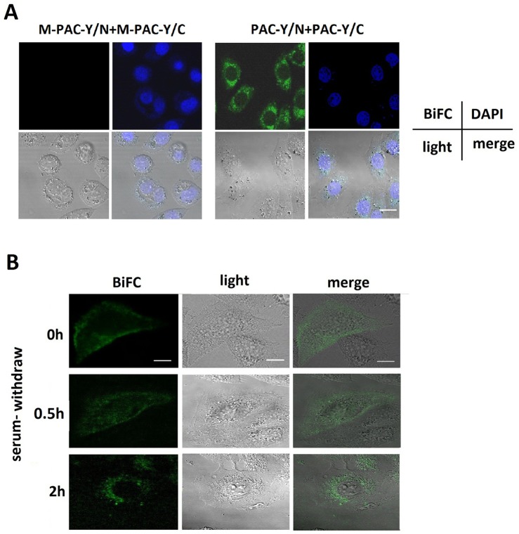
Figure 7. The endocytosis of PAC1 dimers using BiFC during serum withdrawal.
(A) The fluorescence confocal microscopic images of BiFC signals 2 h after serum withdrawal. It was shown that after the nuclear staining with DAPI in CHO cells transfected with PAC-Y/N+PAC-Y/C, the BiFC signals (YFP fluorescence reproduced by the dimerization), representing the PAC1 dimers, were mostly located into the cells and close to the nucleus 2 h after serum withdrawal, while CHO cells transfected with M-PAC-Y/N+M-PAC-Y/C displayed no BiFC signals. Bar, 10 µm. (B) The fluorescence confocal microscopic images of live CHO cells transfected with PAC-Y/N+PAC-Y/C submitted to serum withdrawal. At the beginning of the serum withdrawal, the BiFC signals, representing the PAC1 dimers, were mostly located on or near plasma membranes. Then, at 0.5 h after serum withdrawal, the BiFC signals trafficked into the cells in some type of vehicles, and at 2 h after serum withdrawal, most of the BiFC signals were inside the cells and aggregated around the nucleus. Bar, 5 µm.

