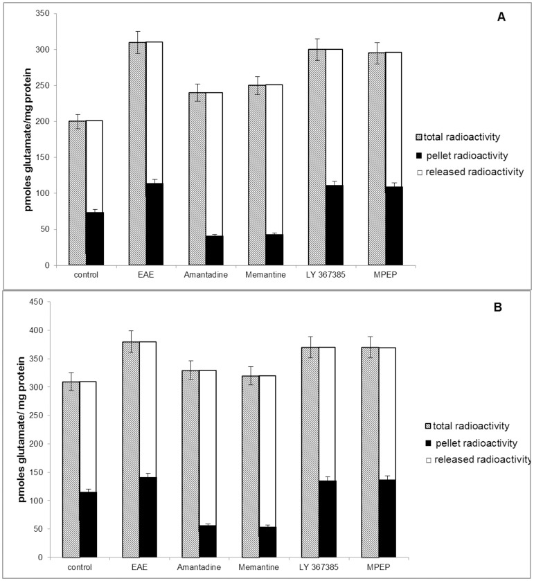
Figure 2. Glutamate release from synaptosomes (A) and GPV fraction (B) obtained from control, EAE, EAE+amantadine, EAE+memantine, EAE+LY367385, and EAE+MPEP rat brains during the acute phase of the disease (12 d.p.i.).
Bars present total radioactivity in pellet (KCl no added), radioactivity remaining in pellet after depolarization by KCl (pellet radioactivity), and released of [3H] glutamate radioactivity from pellet after depolarization by KCl. Membranes were depolarized with 50 mM KCl at a maximum of the uptake curves (6 min), and radioactivity was assayed after 6 min. Results represent the means ± SD from five separate experiments performed in triplicate; * P<0.05 significantly different from the spontaneous release control group; # P<0.05 vs. EAE rats not subjected to therapy (Student's t-test).

