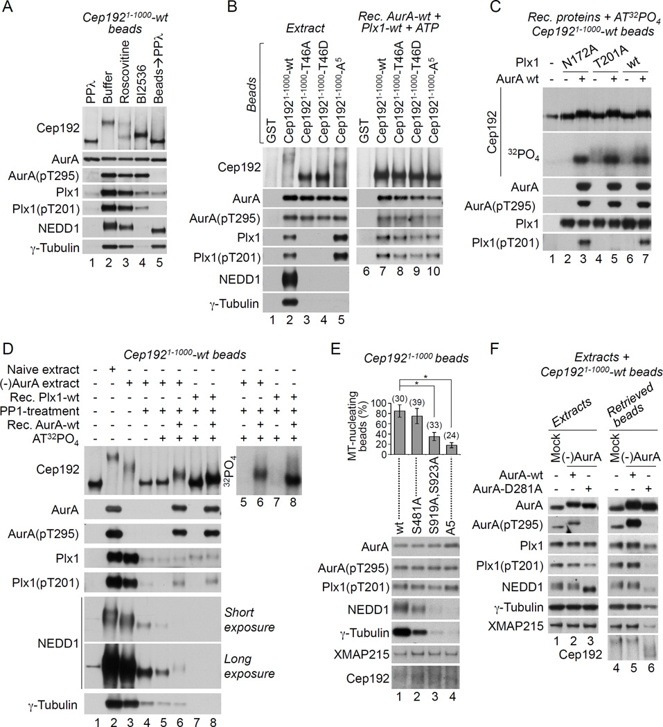
Figure 3. Cep192 organizes AurA and Plx1 in a kinase cascade that drives γ-TuRC recruitment.
(A) W-blot of Cep1921–1000-wt beads incubated in M-phase extracts supplemented as indicated (lanes 1–4). Lane 5: the beads in lane 2 were treated with PPλ and washed in XB 0.1% Tween 20.
(B) W-blot of beads pre-loaded with the indicated GST-proteins and incubated in M-phase extracts (left panel) or in XB supplemented with ATP and recombinant AurA-wt and Plx1-wt proteins (right panel).
(C) W-blot and autoradiogram (32PO4) of Cep1921–1000-wt beads incubated with AT32PO4 in the absence/presence of recombinant AurA-wt and of recombinant Plx1-wt or its enzymatycally inactive (N172A) or non-phosphorylatable (T201A) counterparts. Note that diffuse phosphorylated Cep192 forms are generated by Plx1-wt and Plx1-T201A but not by Plx1-N172A (32PO4).
(D) W-blot (left panel) and autoradiogram (right panel) of Cep1921–1000-wt beads incubated in XB (lane 1), in naïve (lane 2) or AurA-depleted (lanes 3–6) M-phase extracts, or in XB supplemented with recombinant Plx1-wt (lanes 7, 8). In lane 4, the retrieved beads were treated with PP1. In lanes 5–8, the beads were retrieved, treated with PP1, washed, and incubated in the presence of AT32PO4 without/with recombinant AurA-wt.
(E) W-blot of beads pre-loaded with Cep1921–1000-wt or with its mutants lacking one (S481), two (S919 and S923), or five (A5) γ-TuRC-binding serines after the incubation in M-phase extract. The graph shows the mean percentage of bead-induced MT asters +/− SD. The number of beads analyzed is in parentheses. *P<0.001.
(F) W-blots of mock-treated and AurA-immunodepleted extracts supplemented with recombinant AurA-wt or AurA-D281A and with Cep1921–1000-wt beads (left panel) and of Cep1921–1000-wt beads retrieved from these extracts (right panel).
See also Figure S5.

