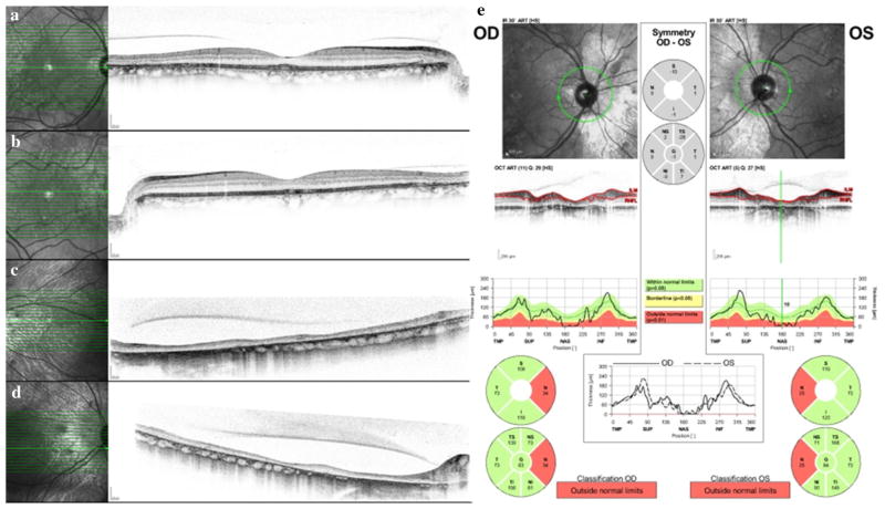
Fig. 2.
Spectral mode OCT images through central fovea in right eye (a) and left eye (b). Note retinal thinning and loss of the outer retinal ellipsoid line temporally (arrows) corresponding to areas of hyper-autofluorescence. Spectral mode OCT images nasal to the optic disks in the right eye (c) and left eye (d). The retinas are severely thinned, with loss of the layered retinal architecture. The choriocapillaris is absent, and adjacent to the optic disks, the larger choroidal vessels are effaced as well. e Nerve fiber layer OCT shows bilateral nasal nerve fiber layer thinning

