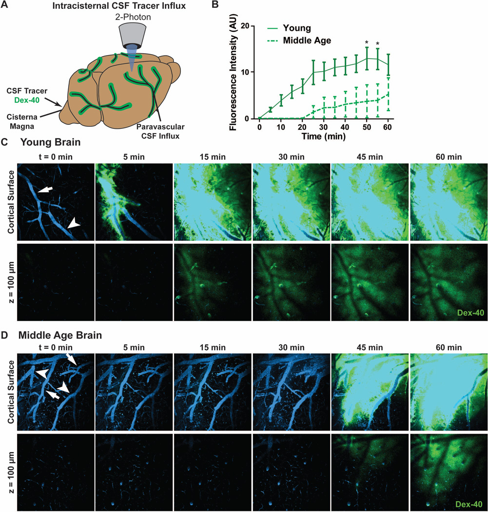
Figure 2. In vivo 2-photon imaging reveals suppressed paravascular glymphatic CSF recirculation in aging mice.
(A) Paravascular CSF tracer penetration into the mouse cortex was evaluated by in vivo 2-photon microscopy after intracisternal injection of FITC-conjugated dextran (40 kD, Dex-40). (B) Quantification of CSF tracer penetration 100 µm below the cortical surface shows impaired paravascular penetration in middle age compared to young cortex (*P<0.05, 2-way repeated measures ANOVA; n = 4 animals per group). (C-D) Serial in vivo 2-photon imaging at the cortical surface and 100 µm below the cortical surface after intracisternal Dex-40 injection. Cerebral vasculature is visualized by intra-arterial injection of Texas Red-conjugated dextran (70 kD, Dex-70, blue); surface arteries (arrows) and veins (arrowheads) are defined morphologically. Compared to the young brain (C), CSF tracer movement along the surface and penetrating arteries and into the surrounding interstitium is markedly slowed in the middle age brain (D).

