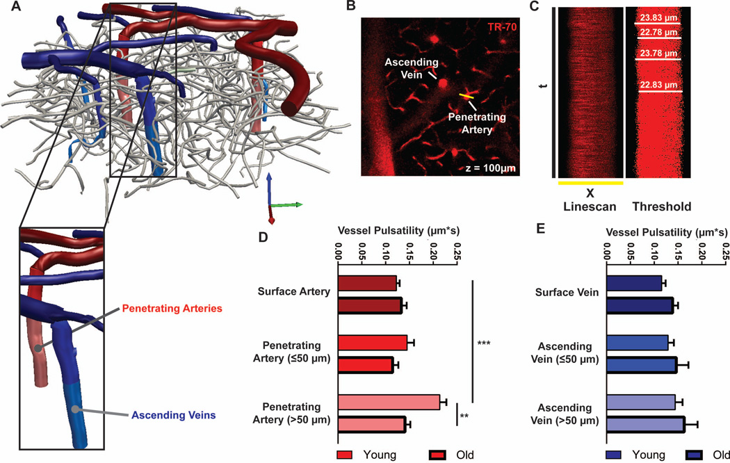
Figure 3. Vascular pulsatility is suppressed in the penetrating arteries of the aging brain.
Pulsatility of the vascular wall was evaluated by in vivo 2-photon microscopy through thin-skull cranial window after intra-arterial injection of Texas Red-conjugated dextran (MW 70kD, dex-70). (A) 3D reconstruction of the cerebrovascular tree visible through cranial window reveals penetrating arteries (red), ascending veins (blue) and intervening capillary bed (gray). (B) Between z = 0 and 150 µm below the cortical surface, high-frequency orthogonal linescans were generated across surface and penetrating arteries and surface and ascending veins. (C) Representative raw x-t scans were thresholded to improve edge detection, and the luminal diameter was measured over time. (D-E) Vascular pulsatility was measured from penetrating arteries and ascending veins. In the young brain, arterial pulsatility was significantly greater than venous pulsatility (*P<0.05, 2-way ANOVA; n = 8–20 vessels from 4 animals per group). In the old brain, arterial pulsatility in the penetrating arteries was significantly reduced compared to the young brain (*P<0.05, Young vs. Old). No age-related differences in venous pulsatility were observed.

