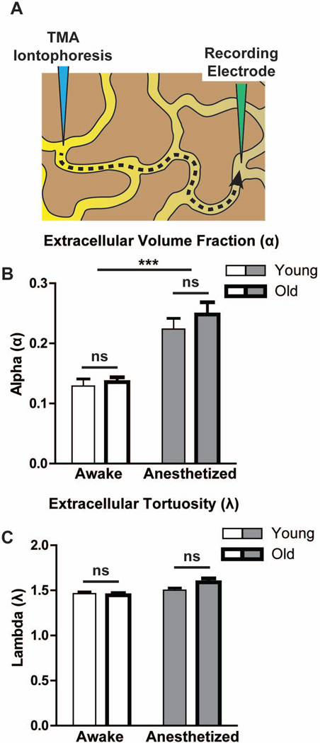
Figure 4. Interstitial volume does not change as a function of aging.
(A) Extracellular volume fraction (α) and tortuosity (λ) were evaluated by the in vivo TMA micro-iontophoresis method. TMA is introduced by an iontophoresis electrode and detected by a second recording electrode. Changes in the extracellular volume fraction or the tortuosity of the extracellular space are reflected in the kinetics of the measured TMA concentrations (described in detail in(18)). (B) In the cortex of either awake or anesthetized mice, extracellular volume fraction did not significantly differ between young and old animals. Both young and old brains exhibited a significant and comparable enlargement of the extracellular space in the anesthetized state (***P<0.001, Awake vs. Anesthetized; 2-Way ANOVA; n = 9–20 per group). (C) Extracellular tortuosity did not differ significantly between young and old animals.

