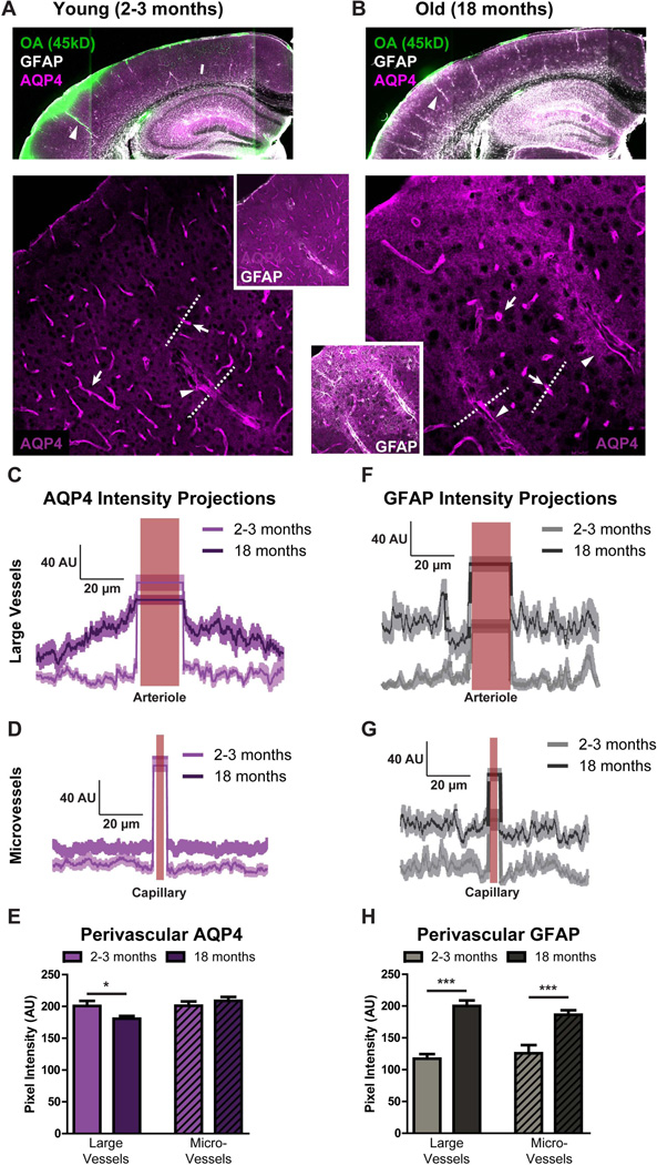
Figure 5. Perivascular AQP4 polarization surrounding penetrating arteries is lost in the aging brain.
Changes in AQP4 localization, GFAP expression and paravascular CSF recirculation were evaluated by immunofluorescence double-labeling. (A-B) Slices from brains fixed 30 min after intracisternal injection of fluorescent CSF tracer ovalbumin-conjugated ALEXA-647 (MW 45kD, OA-45) were imaged by confocal microscopy and show that compared to the young cortex (A), AQP4 localization becomes more disperse in the old brain while GFAP expression increases and CSF tracer penetration is slowed (B). AQP4 and GFAP immunofluorescence were evaluated in linear regions of interest (dashed lines) extending outward from penetrating cerebral arterioles (arrowheads, C, F) or outward from cerebral capillaries (arrows, D, G). (C-D, F-G) Compared to the young brain, and GFAP expression were increased surrounding blood vessels in the aging brain (***P<0.001, young vs. old; repeated measures 2-way ANOVA; n = 11–18 vessels from 4 animals per group). Changes in AQP4 expression were greater surrounding penetrating arterioles (C) than cortical capillaries (B), with marked upregulation of AQP4 in tissue surrounding penetrating vessels. Measurement of perivascular AQP4 (E) and GFAP (H) expression showed that AQP4 is downregulated in perivascular domains surrounding penetrating arterioles, but not around capillaries (*P< 0.05; un-paired t-test, n = 11–18 vessels from 4 animals per group) while perivascular GFAP expression was similarly upregulated surrounding both vessel types (***P<0.001; un-paired t-test).

