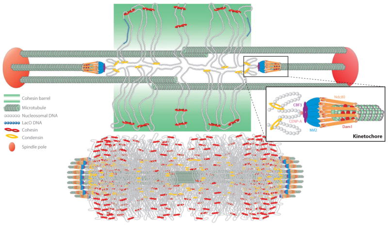
Figure 2.
Organization of pericentric chromatin and cohesin in metaphase in budding yeast. (Top) The yeast segregation apparatus is a composite structure of the kinetochore and interpolar microtubules ( green), the spindle pole body (large red sphere), and pericentric chromatin loops (DNA strands shown as strings of nucleosomes; gray). Centromere DNA (CENP-A nucleosome; pink) is attached to microtubule plus-ends via the kinetochore (orange barbells surrounding the microtubule plus-end). Kinetochore components (right) include Ndc80 (orange barbells), the Dam1 complex (small red spheres interleaved with Ndc80), the Mif2 complex (blue rods), and the DNA-binding complex CBF3 ( purple ovals). Cohesin (red ) and condensin ( yellow) are enriched in the pericentromere and surround the central spindle. Cohesin is radially displaced from the spindle axis, whereas condensin is proximal to the spindle axis (142, 143). One pair of sister chromatids is shown for simplicity. Sister chromatids occupy the left and right half-spindle, respectively. Sister chromatids at the mid-spindle position (perpendicular to the spindle axis) are held via cohesin rings (red ). As sister DNA strands become proximal to the spindle microtubules, they adopt a cruciform-like DNA configuration, with intermolecular sister pairing midway between the spindle poles and intramolecular pairing to the left and right. Condensin rings along the spindle axis contribute to formation of intramolecular loops shown perpendicular to the spindle axis, proximal to the left and right kinetochore. Lac operator DNA (nucleosomes; blue) is radially displaced from the spindle axis when visualized with LacI-GFP (2), similar in dimension to the cohesin barrel. (Bottom) The pericentric chromatin from all 16 chromosomes is shown together with kinetochore microtubules in metaphase. The amount of DNA is drawn to scale and represents the region of DNA from all 16 centromeres that is enriched in the SMC (structural maintenance of chromosome) protein complexes cohesin and condensin. Adapted from figure 4 of Reference 62.

