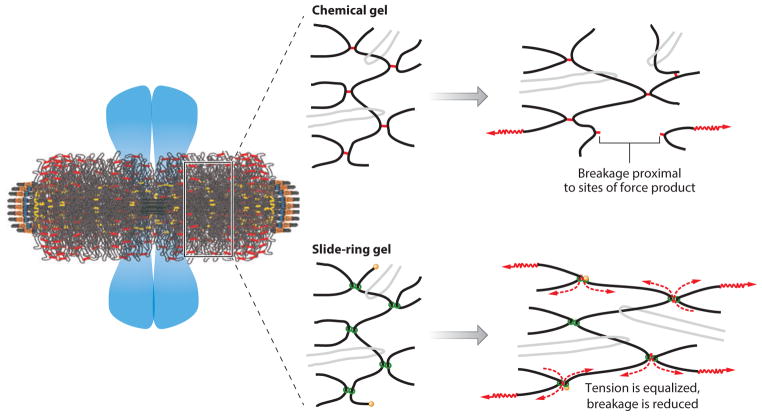
Figure 3.
Strategies for cross-linking centromere DNA in the primary constriction of metaphase chromosomes. (Left) The organization of centromeric heterochromatin relative to chromosome arms in mitosis. The pericentric chromatin from 16 chromosomes surrounding the spindle axis (see key in Figure 2). Sister chromatid arms relative to centromeric heterochromatin are indicated by the blue masses. Pericentric chromatin functions as a contractile element in the spindle, counteracting extensional forces from microtubule-based motor proteins to achieve force balance in metaphase. (Middle) Various ways to generate chromatin cross-links. In a chemical gel (top), inflexible links (red ) connect DNA strands (black). In a slide-ring gel (bottom), molecular pulleys ( green), through which DNA can slide, connect DNA strands. The light gray strands indicate entanglements in which DNA strands are catenated. (Right, top) When force is exerted on DNA strands in a chemical gel, strain is concentrated on inflexible linkages, resulting in rupture. (Right, bottom) When force is exerted on DNA strains that can slip through molecular linkages, tension is distributed throughout the network, equalizing strain among all links, reducing rupture (57, 116).

