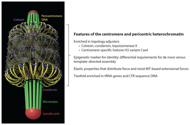
Figure 5.
Emergent properties of the centromere. (Left) The yeast segregation apparatus, shown in the vertical position, serves as a model for multiple kinetochore-microtubule attachment-site centromeres found in other organisms. The kinetochore microtubules ( green) can be seen at the top and bottom, emanating from the spindle pole body (red ). The pericentromere DNA is organized as a network of loops ( yellow) radially arranged around the spindle axis. Cohesin (blue) and condensin ( purple) are enriched in the pericentromere and surround the central spindle. Abbreviations: LTR, long terminal repeat; MT, microtubule. Adapted from figure 6 in Reference 63.

