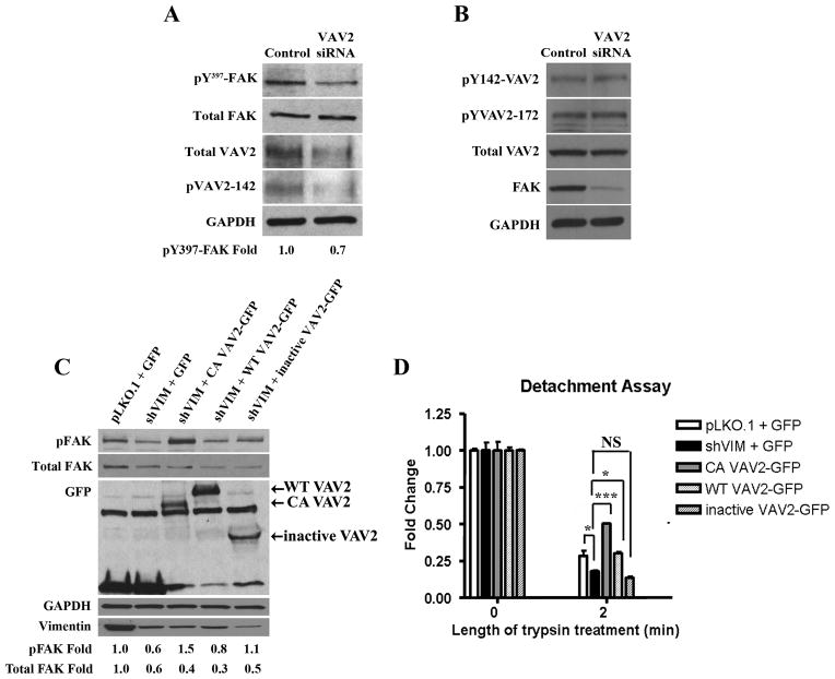
Figure 6. VAV2 regulates vimentin dependent FAK activation and cell adhesion.
(A) Lysates from H1299 cells transfected with VAV2 or control siRNA were analyzed by Western blotting. pY397-FAK levels were reduced upon VAV2 depletion. A representative western blot out of multiple independent experiments is shown. Fold changes were determined by densitometry. (B) Lysates from H1299 cells transfected with FAK or control siRNA were analyzed by western blotting. There was no change in pY142-VAV2, pY172-VAV2 or total VAV2 levels upon FAK depletion. (C) Western blot of H1299 pLKO.1 cells transfected with GFP and H1299 shVIM cells transfected with GFP, CA VAV2-GFP, WT VAV2 or inactive VAV2 shows that CA VAV2-GFP expression rescued the pY397-FAK defect seen in GFP transfected shVIM cells. A representative western blot out of multiple independent experiments is shown. Fold changes were determined by densitometry. (D) A detachment assay with H1299 pLKO.1 cells transfected with GFP and H1299 shVIM cells transfected with GFP, CA VAV2-GFP, WT VAV2 or inactive VAV2 shows that CA VAV2-GFP but not inactive VAV2-GFP expression rescued the cell adhesion defect seen in GFP transfected shVIM cells. * p < 0.05, *** p < 0.001.

