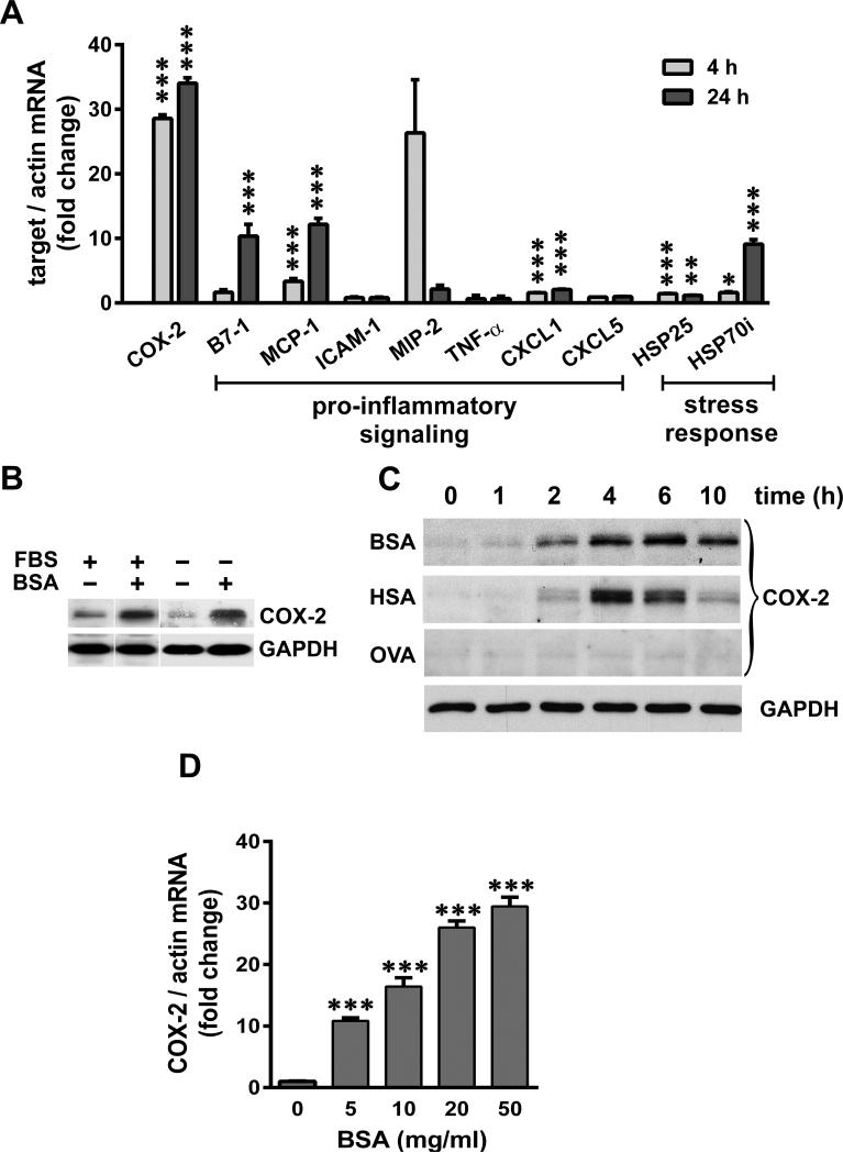
Figure 2.
SA induces COX-2, pro-inflammatory and stress genes in cultured podocytes. A) Serum-starved podocytes were exposed to 40 mg/ml BSA for 4 h and 24 h, and relative mRNA levels of COX-2, pro-inflammatory and stress response genes were measured by qRT-PCR and normalized to β-actin (*P<0.05, **P<0.01 and ***P<0.001 versus time matched control, determined by t test). B) Podocytes were maintained in culture medium containing FBS or serum-starved O/N, and exposed to 40 mg/ml of BSA for 4 h and subjected to immuno-blot analysis for COX-2 and GAPDH. C) Serum-starved podocytes were exposed to 40 mg/ml BSA, human serum albumin (HSA) and Ovalbumin (OVA), and harvested at indicated time points. Protein extracts were analyzed for COX-2 and GAPDH (shown as loading control for a representative blot). D) Serum-starved podocytes were exposed for 4 h to increasing concentrations of BSA as indicated, and mRNA levels of COX-2 were measured by qRT-PCR and normalized to β-actin (***P<0.001 versus control, determined by t test).

